Background
Solute carrier (SLC) proteins are the largest membrane transport group in humans, playing a vital role in transporting nutrients, neurotransmitters, ions, and drugs across cellular membranes. They are crucial for the regulation of cell signaling and the organization of cellular organelles. Despite their importance, SLC proteins remain a highly understudied class of proteins. We offer specialized services for the expression of solute carrier (SLC) proteins, utilizing virus-like particles (VLPs) or nanodiscs to ensure optimal conformation and activity. These advanced expression systems are designed to support a variety of applications, including immunization, phage display, yeast display, ELISA, and SPR.
Custom Protein Workflow
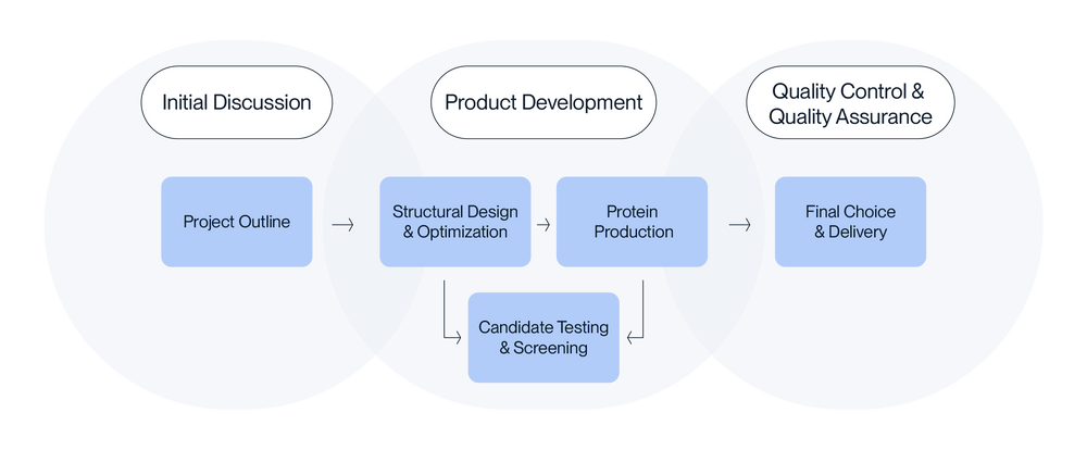
Request Quote:
Service Overview
Product Formats
Virus-like-particle (VLP):
We can display SLC proteins on Virus-like Particles (VLPs), enhancing their utility for various research and therapeutic purposes. VLPs have versatile applications, including immunization and antibody drug discovery, phage display screening, yeast display screening, PK/PD studies, and analytical tests such as ELISA and SPR/BLI. Additionally, our team can customize your VLP to meet your experimental requirements, including immunization guides, in vivo biotinylation, fluorescent labeling, and FACS-compatible VLPs.
Nanodisc:
Our copolymer nanodisc expression can display SLC proteins with detergent-free extraction, providing a versatile tool for various research applications. Nanodiscs are utilized in yeast display screening, in vitro functional assays, PK/PD studies, and analytical tests such as ELISA and SPR/BLI. Additionally, we can customize the nanodisc for your research, including in vivo biotinylation, and fluorescent labeling.
Service Features
6 weeks turnaround time

HEK293 Expression

Full-Length Protein

Customized Proteins
Request Quote
Case Studies
Our team has successfully expressed a comprehensive list of full-length SLC family transporters, including SLC1A4, SLC1A5, SLC7A1, SLC15A4, SLC13A5, SLC16A1, SLC16A3, SLC34A2, SLC40A1, SLC44A4, and SLC59A1.
FITC-Equivalent SLC7A1 VLP
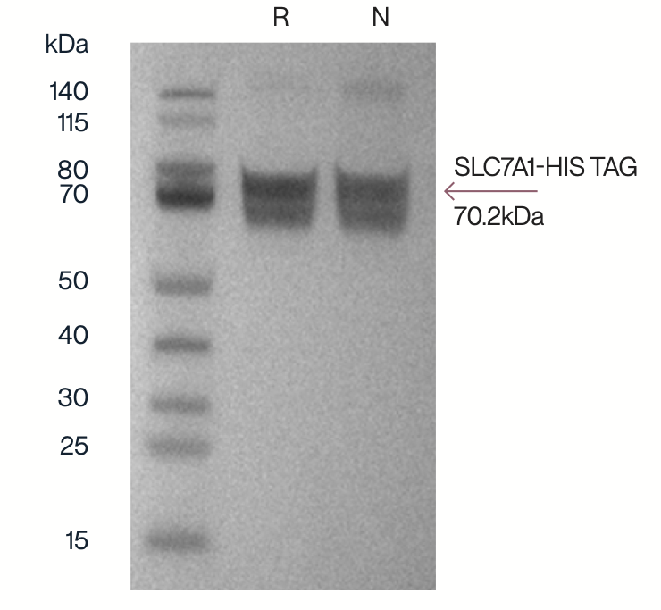
Figure 1. Western blot of full-length SLC7A1 membrane protein with C terminus His tag (70.2kDa) displayed on envelope VLP.
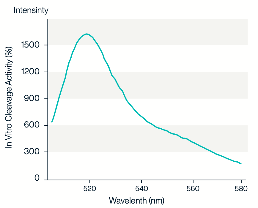
Figure 2. A fluorescent scan of FITC-equivalent SLC7A1 VLP was performed on a Varioskan LUX plate reader (Thermo Fisher). The VLP sample was placed in a 96-well Elisa microplate and measured at 25°C. The excitation wavelength was 488 nm, the emission wavelength was 510 nm, and the measurement range was 505-580 nm.
SLC13A5 Nanodisc
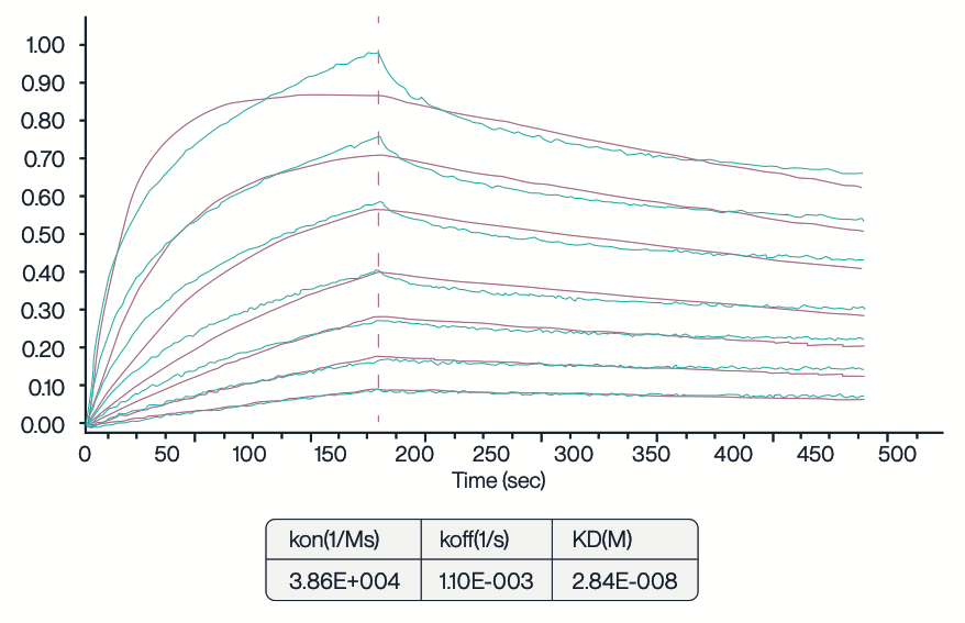
Figure 3. Full-length SLC13A5 nanodisc was tested using (biolayer interferometry). The His-tagged SLC13A5 nanodisc was immobilized on an Anti-His chip, and the binding interaction was analyzed using the 11H7B8 antibody at varying concentrations: 1023.33, 511.67, 255.83, 127.92, 63.96, 31.98, and 15.99 nM. The KD value of 28.4 nM indicates a high affinity of the SLC13A5 nanodisc for the 11H7B8 antibody.
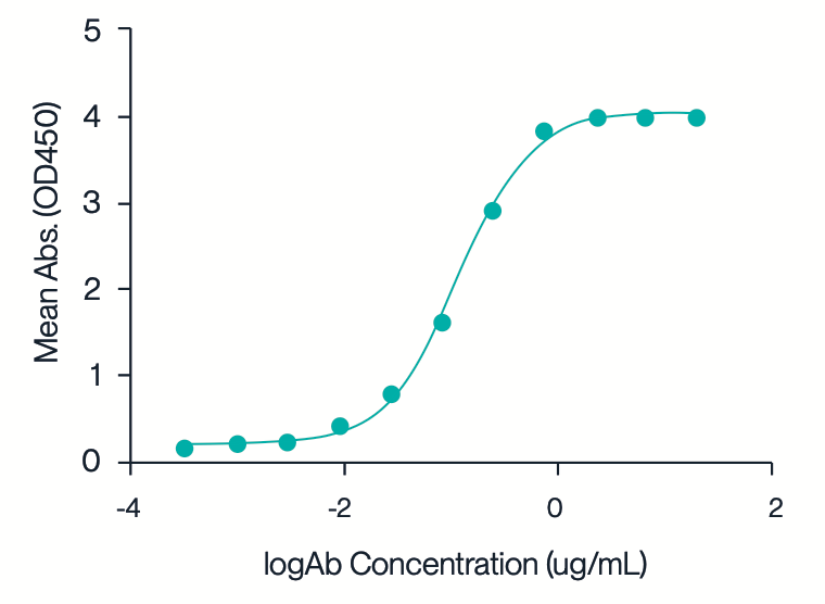
Figure 4. SLC13A5 nanodisc was tested via ELISA using 11H7B8 antibody. The plate was coated with human SLC13A5 nanodisc at 5 µg/mL, and the binding interaction was assessed using a colorimetric assay. The mean absorbance (OD450) was measured across varying concentrations of the antibody, resulting in an EC50 value of 0.14 µg/mL.
SLC13A5 Cell Expression
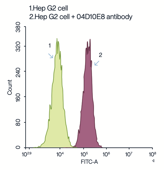
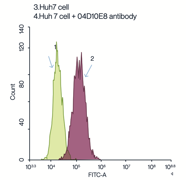
Figure 5. HepG2 and Huh7 cell lines overexpressing SLC13A5 were used for FACS. HepG2 and Huh7 cell lines (2E+06 cells/mL) were incubated with 10 nM recombinant 04D10E8 antibody. Following incubation, cells were labeled with an anti-mouse FC-FITC secondary antibody. The green peaks (1 and 3) represent the autofluorescence of untreated HepG2 and Huh7 cells, respectively. The red peaks (2 and 4) show the fluorescence intensity of HepG2 and Huh7 cells bound with the 04D10E8 antibody. This data demonstrates the binding specificity and expression level of the SLC13A5 membrane protein in these cell lines.
SLC Protein Request Form:

VLP & Nanodisc Transmembrane Proteins

SPR Analysis Services

Custom VLP or Nanodisc


