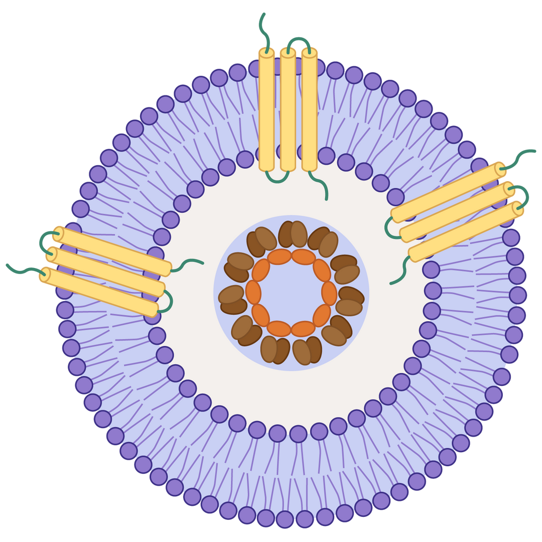
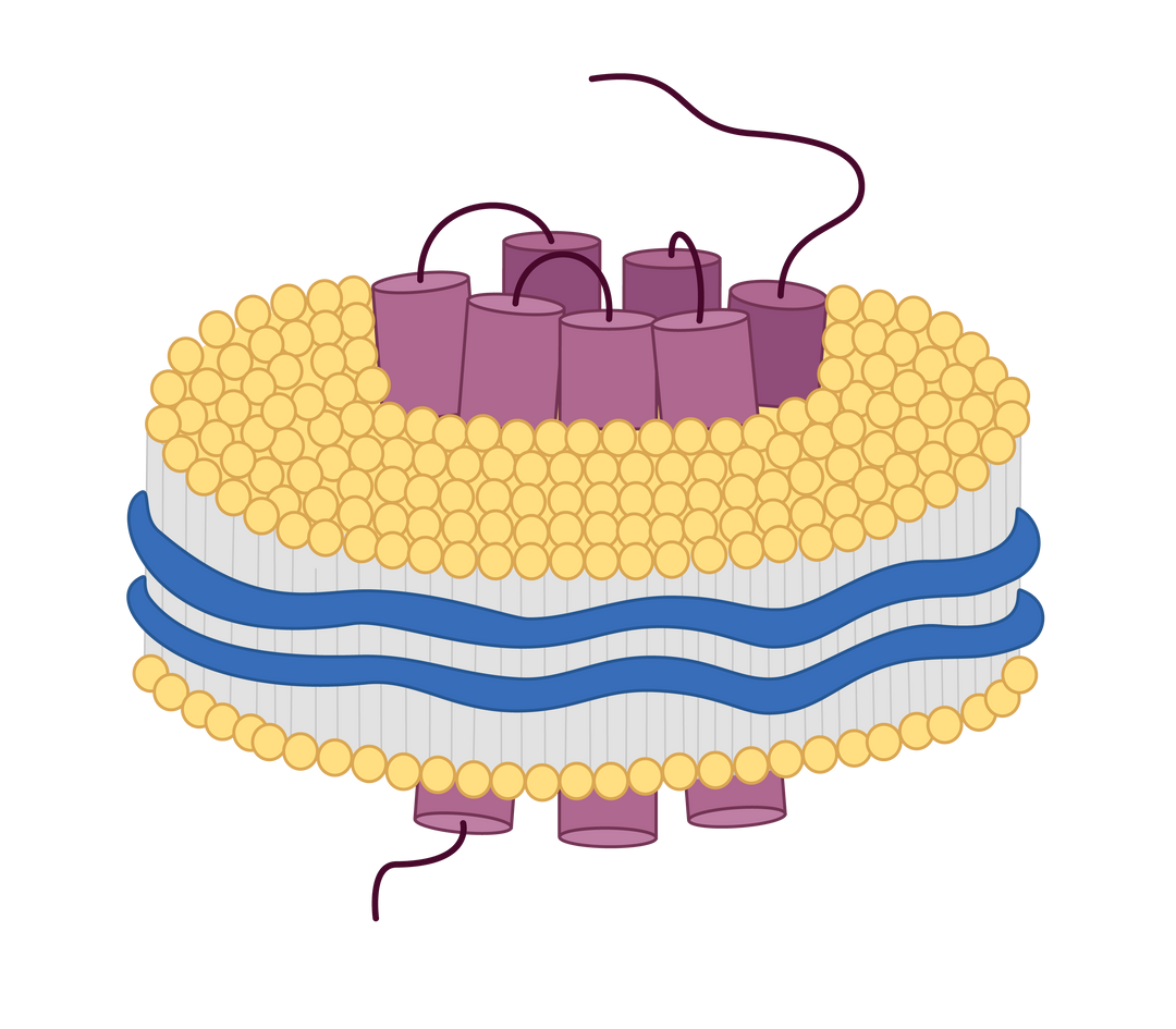
How the VLP displays transmembrane proteins.
Advantages of displaying membrane proteins on VLPs
Native-like Conformation: VLPs provide a lipid bilayer environment that helps transmembrane proteins maintain their correct folding, topology, and conformation, closely mimicking their native state on the cell membrane.
Enhanced Immunogenicity: The repetitive, multivalent display of antigens on VLPs significantly boosts immune responses compared to soluble or monomeric proteins, making them ideal for vaccine and antibody development.
Stabilization of Difficult Targets: Transmembrane proteins are often unstable or insoluble when isolated. VLPs offer structural support, increasing protein stability and solubility during purification and immunization.
Improved Antibody Generation: Presenting transmembrane proteins on VLPs enhances the likelihood of generating antibodies against conformational epitopes, which are often lost in linear peptide or denatured formats.
No Genetic Material = High Safety: VLPs are non-infectious because they lack viral genetic material, offering a safe platform for research and therapeutic development.
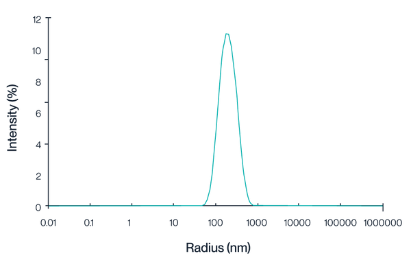
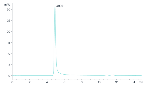
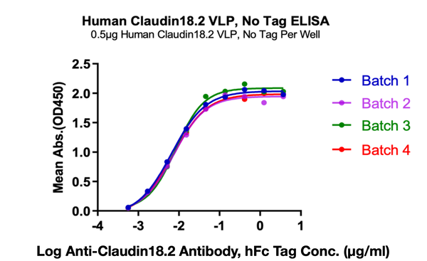
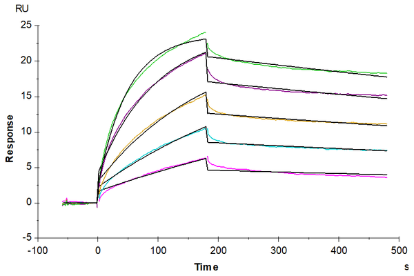
Product Validation
Claudin 18.2 is a tetraspanin protein family member of critical therapeutic potential for gastric and esophageal adenocarcinomas. Despite interest in drug development targeting Claudin 18.2, technical challenges in expressing high-quality Claudin 18.2 protein have significantly limited the progress of Claudin 18.2-targeted drug discovery and development. By leveraging virus-like particles, we have become the first company to successfully produce the commercially available, full-length human Claudin 18.2 protein in soluble format. Our consistent activity and purity testing results demonstrate the functional integrity and bioactivity of our VLP-displayed
protein platform.