


About Nanospheres™
Nanospheres™ are a novel membrane display system engineered to present transmembrane proteins in a highly controlled and purified manner. Built from specialized nanodiscs, Nanospheres™ are artificial membrane spheres that deliver a highly organized and concentrated membrane environment—unlocking new possibilities for applications in antibody immunization, screening, and analytical assays. They can also complement capture and detection workflows that use streptavidin-coated beads for isolating biotinylated targets.
Nanospheres™ are designed to:
→ Mimic natural membrane surfaces
→ Correctly orient proteins with the extracellular domains outward
→ Support multipass transmembrane protein stability
→ Provide highly-dense and highly-pure membrane protein display
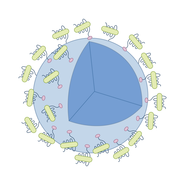
Nanospheres™ are highly-pure, full-length transmembrane protein nanodiscs densely bound to a bead or resin.
Assembly & Structure
Nanospheres™ are engineered by immobilizing purified nanodiscs—each embedded with a target transmembrane protein—onto beads or resin. This modular system ensures a dense, directional, and highly controlled membrane protein display.
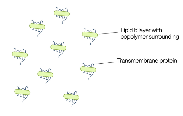
1. Mammalian Expression & Affinity Purification.
The desired multipass transmembrane protein is recombinantly expressed in mammalian cells and incorporated into nanodiscs composed of a lipid bilayer stabilized by copolymers or scaffold proteins. The nanodiscs are purified using affinity chromatography to ensure all nanodics contain the target protein.
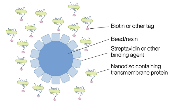
2. Biotinylation or Tagging.
The purified nanodiscs are functionalized at the C-terminus with biotin to enable site-specific attachment while preserving extracellular domain orientation.
3. Conjugation to Beads or Resin.
Tagged nanodiscs are bound to streptavidin-coated or Ni-NTA–coated beads/resins via high-affinity interactions (biotin-streptavidin). This step forms the spherical nanosphere structure.
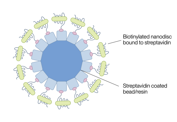
4. Final Format: Oriented, High-Density Display of Target Protein
The resulting Nanospheres™ present the target transmembrane protein in a native-like orientation and membrane context—ideal for downstream assays such as antibody screening, flow cytometry, and ligand-binding studies.
Advantages of Nanospheres™

High Purity
Nanospheres™ greatly reduce background noise by displaying only the target transmembrane proteins, omitting unwanted host membrane proteins found in VLPs.

Dense Target Protein Display
Each Nanosphere™ is densely packed with your protein of interest, providing 36 to 600 times more target membrane proteins compared to VLPs.

Compatible with Toxic Proteins
Nanospheres™ bypass the limitations of cell-based systems. Toxic or hard-to-express transmembrane proteins can be densely incorporated into Nanospheres™, unlike VLPs.
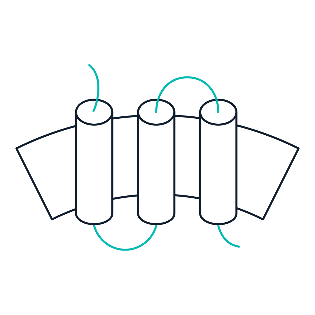
Controlled Orientation of Extracellular Domain
The nanodiscs are immobilized on the Nanosphere™ via the C-termini, ensuring correct extracellular domain orientation—preserving native-like conformation and functionality.
1. Cell-Free Alternative for Flow Cytometry
Nanospheres™ act as a cell-free alternative to live cells for flow cytometry experiments when certain live cell strains are difficult to maintain or with high variability in performance. The consistent, well-controlled display of target transmembrane proteins can significantly reduce variability commonly seen in cell-based assays, increasing data reproducibility.
2. Immunization for Antibody Discovery
Nanospheres™ serves as an effective immunogen for antibody generation, particularly for difficult-to-express multipass transmembrane proteins. The high-density display of purified target proteins enhances immune responses, leading to stronger immunization performance and diverse antibody pool generation, which is critical for downstream monoclonal antibody screening and lead selection.
3. Drug Discovery & High-Throughput Screening (HTS)
Nanospheres™ provide a stable and scalable platform for screening small molecules or biologics that interact with membrane proteins. This is useful for identifying inhibitors or activators of G protein-coupled receptors (GPCRs), ion channels, and transporters, etc, which are key drug targets.
4. Ligand-Binding Assays
Nanospheres™ can be used in ligand binding assays such as SPR and ELISA to detect interactions between membrane proteins and ligands or antibodies. The controlled protein orientation improves assay reproducibility and sensitivity compared to traditional membrane fractions, suggesting broad applications in PK/PD studies, tox studies, ligand identification, or general protein-protein interaction studies.
Product Validation Data
The following flow cytometry data demonstrate the functional presentation and specificity of membrane proteins displayed on Nanospheres™. In both unlabeled and fluorescent formats, Nanospheres™ show strong and specific binding to target antibodies—confirming proper protein orientation, structural integrity, and surface accessibility. These results validate the use of Nanospheres™ in binding assays, cell sorting, and immunotherapeutic screening applications.
Example 1: Nanosphere™ for Cell-Free Flow Cytometry
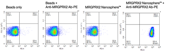
Representative dot plots showing PE signal (x-axis) versus forward scatter (y-axis) for four experimental conditions. (1) Beads only, (2) Beads incubated with anti-MRGPRX2-PE antibody, (3) Beads conjugated with MRGPRX2 Nanosphere™, and (4) MRGPRX2 Nanosphere™-conjugated beads incubated with anti-MRGPRX2-PE antibody. Only the final condition shows a strong PE-positive population (Q2-4 quadrant, 99.98%), indicating specific binding of the anti-MRGPRX2 antibody to the correctly oriented, functional MRGPRX2 displayed on the Nanosphere™. These data validate the use of MRGPRX2 Nanospheres™ for cell-free flow cytometry-based detection and sorting applications.
Example 2: Fluorescent Nanosphere™ for Cell-Free Flow Cytometry
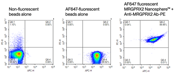
Dot plots display PE fluorescence (y-axis) and APC (AF647) fluorescence (x-axis) across three conditions: (1) Non-fluorescent beads alone, (2) AF647-labeled beads alone, and (3) AF647-labeled MRGPRX2 Nanospheres™ incubated with anti-MRGPRX2-PE antibody. Only the third condition exhibits a distinct double-positive population (Q6-2 quadrant, 99.95%), demonstrating specific binding of the antibody to MRGPRX2 and successful dual fluorescence labeling. These results confirm the utility of fluorescent Nanospheres™ for cell-free, multiplexed flow cytometry assays and targeted sorting applications.
Request Quote:

60 Hickory Drive
Waltham, MA 02451
United States
Nanosphere™ - Novel Membrane Protein Display Platform | KACTUS