KLK5/7: Emerging Targets for Inflammatory Skin Diseases
By Natasha Slepak
The epidermis, as the first barrier, prevents water loss while resisting harmful substance invasions. KLKs, including KLK5, KLK7, and KLK14, are co-expressed in the stratum corneum and upper granular layer of normal human epidermis and associated hair follicle–sebaceous gland units. By promoting desquamation and keratinocyte proliferation, they directly regulate skin renewal and barrier thickness, which are critical for maintaining skin barrier function. When KLK activity becomes dysregulated, excessive desquamation occurs, triggering skin inflammations such as Netherton Syndrome (NS) and Atopic Dermatitis (AD). Studies have shown that upregulation of KLK5 and KLK7 is a major factor leading to increased desquamation in NS. Additionally, KLK7 is a key member involved in the pathogenesis of AD [1]. Thus, KLK5 and KLK7 represent emerging therapeutic targets for skin diseases such as NS and AD.
KLK Proteolytic Cascade
The kallikrein-related peptidase family (KLKs) is a group of secreted serine proteases consisting of 15 members (KLK1–15). The KLK genes are tandemly clustered on chromosome 19q13.4, and 13 members (KLK1, 4–15) have been identified in human skin. Among them, KLK5 and KLK7 were the first two KLK proteins that were identified in skin [1]. During keratinocyte differentiation, KLKs are secreted from lamellar granules (LGs) of upper granular keratinocytes into the intercellular space of the stratum corneum (SC). The secreted, inactive precursors (proKLKs) form an activation cascade. ProKLK5 can self-activate and thus serves as the initiator of the cascade, subsequently activating other skin KLKs such as ProKLK7, ProKLK8, and ProKLK14 through prodomain cleavage. Activated KLK14 can, in turn, feedback positively to activate ProKLK5, amplifying the proteolytic activity. The activated KLK5 and KLK7 degrade intercellular adhesion proteins between keratinocytes (including desmoglein-1 (DSG1), desmocollin-1 (DSC1), and corneodesmosin (CDSN)) to promote desquamation and shedding of corneocytes, thereby maintaining skin homeostasis. KLK5, KLK6, and KLK14 may also activate the protease-activated receptor-2 (PAR2) expressed in keratinocytes by cleaving its N-terminal region. This in turn triggers downstream signaling pathways (eg. NF-kB activation), leading to inflammatory cytokine release or modulation of the lipid permeability barrier.

KLK Proteolytic Cascades [2]
The fine-tuned regulation of KLK cascades is primarily mediated by endogenous serine protease inhibitors, such as the lymphoepithelial Kazal-type–related inhibitor (LEKTI), encoded by the SPINK5 gene. LEKTI contains 15 domains and is produced as a precursor that is rapidly cleaved by furin into multiple single- or multi-domain fragments. Among them, D5, D6, D8–D11, and D9–D15 exhibit diverse inhibitory effects on KLK5, KLK7, and KLK14 [3]. Mutations in SPINK5 cause NS, as the absence of LEKTI leads to uncontrolled activation of KLK5 and downstream KLKs (KLK7 and KLK14), resulting in excessive degradation of DSG1, DSC1, and CDSN. This allows allergens, irritants, and bacteria to penetrate the skin, inducing inflammation, pruritus, and barrier dysfunction [2]. In AD lesions, KLK5, KLK7, and KLK14 are also upregulated [4].
Additionally, the acidic skin surface mantle and epidermal pH gradient play crucial roles in regulating KLKs. From the basal layer to the stratum corneum, pH gradually decreases. At neutral pH, KLK5 and KLK7 show optimal activity, and KLK5 binds tightly to LEKTI fragments. As pH decreases, KLK5 dissociates from LEKTI, ensuring proper desquamation in the stratum corneum.

The pH gradient in epidermis [1]
Advances of KLK5/7 Drug Development
Therapeutics targeting KLK5/7 for NS and AD mainly include monoclonal antibodies, bispecific antibodies, and fusion proteins. TRIV-509 is the most advanced candidate (Phase I trial), a monoclonal antibody targeting both KLK5 and KLK7 for AD. In multiple preclinical AD models, Triveni Bio claims that TRIV-509 demonstrated superior efficacy compared with IL-4R inhibitors and showed rapid improvement in AD patient skin biopsy samples [5].

TRIV-509 monoclonal antibody
RO7449135, developed by Genentech, is a humanized bispecific IgG1 antibody that effectively inhibits KLK5 and KLK7 activity. Structural studies revealed that the anti-hKLK5 Fab targets the 170s loop of KLK5, distant from its active site. This binding disrupts the substrate-binding region of KLK5 via an allosteric inhibition mechanism. In vitro, RO7449135 can completely restore the epithelial barrier permeability disrupted by KLK5/7 and counteract the effects of LEKTI deficiency in human keratinocytes.

The binding between Anti-KLK5 Fab and KLK5 [6]
|
Drug Name |
Target |
Indication |
Modality |
Clinical Stage |
Company |
|
TRIV-509 |
KLK5 × KLK7 |
Atopic Dermatitis (AD) |
mAb |
Phase I |
Triveni Bio |
|
BCX-17725 |
KLK5 |
Netherton Syndrome |
Fusion Protein |
Phase I |
BioCryst Pharmaceuticals |
|
DS-2325a |
KLK5 |
Netherton Syndrome |
Fusion Protein |
Phase I |
Daiichi Sankyo |
|
RO7449135 |
KLK5 × KLK7 |
Netherton Syndrome |
BsAb |
Preclinical |
Genentech |
Selected KLK5/7 Drug Candidates against NS and AD
KACTUS High-Quality KLK5/7 Proteins
As emerging therapeutic targets, KLK5 & KLK7 show great potential in the treatment of NS, AD, and other skin diseases. KACTUS provides the active form of KLK5 and KLK7, which can fulfill the unmet needs of antibody drug development against KLK5/7. Each batch undergoes rigorous activity testing, including enzymatic activity before release, making them suitable for applications such as antibody generation, drug screening and immunoassay development.
Product Data Examples:

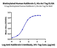
Immobilized Human Kallikrein 5, His Tag at 0.5μg/ml (100μl/Well) on the plate. Dose response curve for Anti-Kallikrein 5 Antibody, hFc Tag with the EC50 of 3.7ng/ml determined by ELISA .(QC Test)

Enzymatic Activity. Measured by its ability to cleave the fluorogenic peptide substrate Boc-VPR-AMC. The specific activity is >400 pmol/min/µg (QC Test).

Immobilized Human Kallikrein 7, No Tag at 0.5μg/ml (100μl/well) on the plate. Dose response curve for Anti-Kallikrein 7 Antibody, hFc Tag with the EC50 of 2.9ng/ml determined by ELISA. (QC Test)

Measured by its ability to cleave the fluorogenic peptide substrate, Mca-RPKPVE-Nval-WRK(Dnp)-NH2. The specific activity is >350 pmol/min/µg. (QC Test)
Product List
|
Catalog Number |
Product Name |
Sequence |
Species |
Tag |
Expression System |
|
Human Kallikrein 5/KLK5 Protein (active form) |
Ile67-Ser293 |
Human |
C-His |
HEK293 |
|
|
Human Kallikrein 5/KLK5 Protein (active form) |
Ile67-Ser293 |
Human |
C-His-Flag |
HEK293 |
|
|
Biotinylated Human Kallikrein 5/KLK5 Protein (active form) |
Ile67-Ser293 |
Human |
C-His-Avi |
HEK293 |
|
|
KLK-CM105 |
Cynomolgus Kallikrein 5/KLK5 Protein (active form) |
Ile65-Ser291 |
Cynomolgus |
C-His |
HEK293 |
|
Mouse Kallikrein 5/KLK5 Protein (active form) |
Gly30-Asn293 |
Mouse |
C-His |
HEK293 |
|
|
Human Kallikrein 7/KLK7 Protein (active form) |
Ile30-Arg253 |
Human |
No Tag |
HEK293 |
|
|
Human Kallikrein 7/KLK7 Protein (active form) |
Ile30-Arg253 |
Human |
C-His |
HEK293 |
|
|
Human Kallikrein 7/KLK7 Protein (pro form) |
Glu23-Arg253 |
Human |
C-His |
HEK293 |
|
|
Human Kallikrein 7/KLK7 Protein (pro form) |
Glu23-Arg253 |
Human |
C-His-Flag |
HEK293 |
|
|
Biotinylated Human Kallikrein 7/KLK7 Protein (pro form) |
Glu23-Arg253 |
Human |
C-His-Avi |
HEK293 |
|
|
KLK-CM107 |
Cynomolgus Kallikrein 7/KLK7 Protein (active form) |
Ile30-Arg253 |
Cynomolgus |
C-His |
HEK293 |
|
Mouse Kallikrein 7/KLK7 Protein (active form) |
Ile26-Arg249 |
Mouse |
C-His |
HEK293 |
References
[1]Di Paolo CT, Diamandis EP, Prassas I. The role of kallikreins in inflammatory skin disorders and their potential as therapeutic targets. Crit Rev Clin Lab Sci. 2021 Jan;58(1):1-16. doi: 10.1080/10408363.2020.1775171. Epub 2020 Jun 22. PMID: 32568598.
[2]Prassas I, Eissa A, Poda G, Diamandis EP. Unleashing the therapeutic potential of human kallikrein-related serine proteases. Nat Rev Drug Discov. 2015 Mar;14(3):183-202. doi: 10.1038/nrd4534. Epub 2015 Feb 20. Erratum in: Nat Rev Drug Discov. 2015 Oct;14(10):732. PMID: 25698643.
[3]Deraison C, Bonnart C, Lopez F, Besson C, Robinson R, Jayakumar A, Wagberg F, Brattsand M, Hachem JP, Leonardsson G, Hovnanian A. LEKTI fragments specifically inhibit KLK5, KLK7, and KLK14 and control desquamation through a pH-dependent interaction. Mol Biol Cell. 2007 Sep;18(9):3607-19. doi: 10.1091/mbc.e07-02-0124. Epub 2007 Jun 27. PMID: 17596512; PMCID: PMC1951746.
[4]Morizane S. The Role of Kallikrein-Related Peptidases in Atopic Dermatitis. Acta Medica Okayama. 2019 Feb;73(1):1-6. DOI: 10.18926/amo/56452. PMID: 30820048.
[4]LB1256 TRIV-509, a dual inhibitor of KLK5 and KLK7, rapidly improves barrier integrity and markers of epidermal differentiation in atopic dermatitis skin explants. Mateer, E. et al.Journal of Investigative Dermatology, Volume 145, Issue 8, S221
[5]Science and Pipeline | Triveni Bio
[6]Chavarria-Smith J, Chiu CPC, Jackman JK, Yin J, Zhang J, Hackney JA, Lin WY, Tyagi T, Sun Y, Tao J, Dunlap D, Morton WD, Ghodge SV, Maun HR, Li H, Hernandez-Barry H, Loyet KM, Chen E, Liu J, Tam C, Yaspan BL, Cai H, Balazs M, Arron JR, Li J, Wittwer AJ, Pappu R, Austin CD, Lee WP, Lazarus RA, Sudhamsu J, Koerber JT, Yi T. Dual antibody inhibition of KLK5 and KLK7 for Netherton syndrome and atopic dermatitis. Sci Transl Med. 2022 Dec 14;14(675):eabp9159. doi: 10.1126/scitranslmed.abp9159. Epub 2022 Dec 14. PMID: 36516271.


